.jpg?width=438&height=298&ext=.jpg)
A new PET/CT (positron emission tomography) exam and a new radiopharmaceutical tracer adds up to patients like Chuck Van Vorst accessing a life altering procedure close to home.
Having part of his pancreas and all of his spleen removed at Mayo Clinic due to pancreatic cancer in 2014, Van Vorst was fortunate to have access to the new radiology exam this year at the Carle Urbana campus. Medical staff discovered an idle neuroendocrine tumor which took only seven incisions to remove.
“I was lucky to stay locally and have the newer test done at Carle,” he said. Van Vorst is no stranger to medical procedures as he once was CEO of Carle Foundation Hospital. He said Carle purchased its first MRI machine when he was CEO and medicine has come so far.
Carle Imaging Manager for Radiology Thorin Mayba said it simply: “This is a game changer for our community.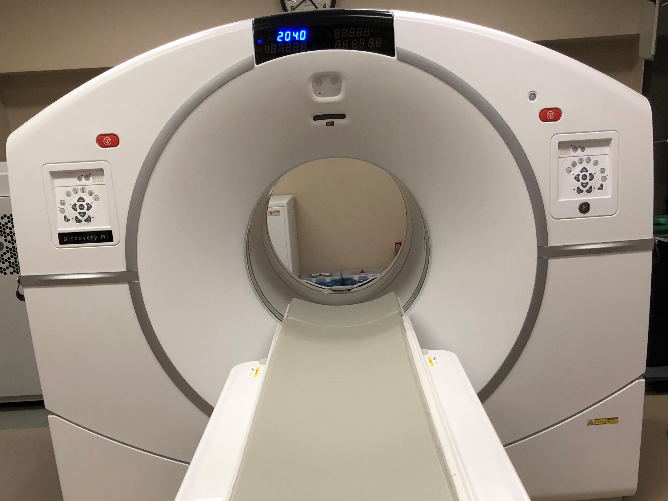 The new scanning equipment and access to the new radiopharmaceutical tracer keeps the availability of the technology in our back yard.”
Brett Yockey, MD, at Carle Foundation Hospital Radiology, was at a conference when he learned of the new tracer. Providing patient access to the PET testing was impossible as the tracer had just a 68 minute half-life and it is a two-hour drive to the closest radio pharmacy that can provide this new tracer. “There was no choice but to send patients to larger hospitals located hours away,” Yockey said.
The new tracer combined with the new PET/CT scanner makes it easier to detect how many neuro endocrine tumors are in a particular patient as the machine scans the entire body of the patient. The tracer also gives technologists more confidence in the clearer images they see.
“We want to make sure our patients have the same access as someone at Mayo,” Yockey said.
Called Copper 64 Detectnet, the tracer has more than a 12 hour half-life. Used in combination with new PET/CT scanning equipment in radiology, neuro-endocrine imaging procedures that used to take multiple days to complete now finish in the same day without needing bowel prep. Imaging time alone with this new procedure takes 30 minutes versus an hour to 90 minutes on multiple days with the old exam.
To learn more about Carle’s Radiology Department, go to https://carle.org/Services/Radiology.
Having part of his pancreas and all of his spleen removed at Mayo Clinic due to pancreatic cancer in 2014, Van Vorst was fortunate to have access to the new radiology exam this year at the Carle Urbana campus. Medical staff discovered an idle neuroendocrine tumor which took only seven incisions to remove.
“I was lucky to stay locally and have the newer test done at Carle,” he said. Van Vorst is no stranger to medical procedures as he once was CEO of Carle Foundation Hospital. He said Carle purchased its first MRI machine when he was CEO and medicine has come so far.
Carle Imaging Manager for Radiology Thorin Mayba said it simply: “This is a game changer for our community.
The new scanning equipment and access to the new radiopharmaceutical tracer keeps the availability of the technology in our back yard.”Brett Yockey, MD, at Carle Foundation Hospital Radiology, was at a conference when he learned of the new tracer. Providing patient access to the PET testing was impossible as the tracer had just a 68 minute half-life and it is a two-hour drive to the closest radio pharmacy that can provide this new tracer. “There was no choice but to send patients to larger hospitals located hours away,” Yockey said.
The new tracer combined with the new PET/CT scanner makes it easier to detect how many neuro endocrine tumors are in a particular patient as the machine scans the entire body of the patient. The tracer also gives technologists more confidence in the clearer images they see.
“We want to make sure our patients have the same access as someone at Mayo,” Yockey said.
Called Copper 64 Detectnet, the tracer has more than a 12 hour half-life. Used in combination with new PET/CT scanning equipment in radiology, neuro-endocrine imaging procedures that used to take multiple days to complete now finish in the same day without needing bowel prep. Imaging time alone with this new procedure takes 30 minutes versus an hour to 90 minutes on multiple days with the old exam.
To learn more about Carle’s Radiology Department, go to https://carle.org/Services/Radiology.
Categories: Culture of Quality, Redefining Healthcare, Community
Tags: CT scanner, pancreatic cancer, radiology