Traditionally, someone in a lab will embed tissue in paraffin, pour dye and view it under a microscope to determine if cancer cells are present.
However, researchers in bioengineering and medicine at Carle are going further and looking at the microenvironment, or molecular chemistry of the cancer cells to understand cancer progression. By measuring the molecular makeup of cancer cells and their effect on the surrounding cells and tissues, that information can provide information to patients as to how quickly they need treatment.
To accomplish this, they want to build a microscope for a clinical setting to change how to measure and view cells.
 “There is no such thing as an insignificant cancer for the patient. However, all cancers do not behave the same,” said Rohit Bhargava, PhD, professor of bioengineering and director of the Cancer Center at Illinois, which is dedicated to cancer-related research and scholarship at the University of Illinois Urbana-Champaign.
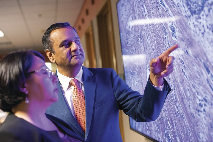
Bhargava and Georgina Cheng, MD, PhD, a cancer surgeon at Carle Cancer Institute Urbana, said their research of labeling unique components of the tumor and its cells through use of infrared-based organizational measurements is predictive of overall survival of colon cancer.
“This is a different way of looking at cancer cells,” Bhargava said. “We asked what is the role of each type of cell in the cancer’s microenvironment and how do the characteristics of each cell relate to the other cells? The spatial organization of those cells are important for prediction of survival for someone with colon cancer, which is one of the deadliest cancers.”
 Dr. Cheng said, “Our goal, at the time of the patient’s cancer surgery, is to provide information about the tumor that can be used to make decisions during surgery and to guide treatment.”

The information gathered from measuring the makeup of each cancer cell can give a patient a clear view of their best choices for treatment, Bhargava said. “The information empowers the patient and it empowers the clinician in providing treatment to a more at-risk group faster.”
Jennifer Eardley, PhD, vice president for Research at Carle Foundation Hospital, said, “The groundbreaking research conducted by Drs. Bhargava and Cheng could lead to major improvements in the way surgeons approach tumor removal and plan follow-up treatment. Having real-time information regarding the spread of cancer in the operating room will help surgeons make better decisions to catch all diseased tissues and save areas that are healthy, which ultimately will improve outcomes for patients.”
The prestigious Science Advances publication of the American Association for the Advancement of Science recently published their work.-
To move forward, they are seeking federal funding in order to build a microscope they’ll use to tell them about each cancer cell composition in a clinical setting.
However, researchers in bioengineering and medicine at Carle are going further and looking at the microenvironment, or molecular chemistry of the cancer cells to understand cancer progression. By measuring the molecular makeup of cancer cells and their effect on the surrounding cells and tissues, that information can provide information to patients as to how quickly they need treatment.
To accomplish this, they want to build a microscope for a clinical setting to change how to measure and view cells.
“There is no such thing as an insignificant cancer for the patient. However, all cancers do not behave the same,” said Rohit Bhargava, PhD, professor of bioengineering and director of the Cancer Center at Illinois, which is dedicated to cancer-related research and scholarship at the University of Illinois Urbana-Champaign.Bhargava and Georgina Cheng, MD, PhD, a cancer surgeon at Carle Cancer Institute Urbana, said their research of labeling unique components of the tumor and its cells through use of infrared-based organizational measurements is predictive of overall survival of colon cancer.
“This is a different way of looking at cancer cells,” Bhargava said. “We asked what is the role of each type of cell
in the cancer’s microenvironment and how do the characteristics of each cell relate to the other cells? The spatial organization of those cells are important for prediction of survival for someone with colon cancer, which is one of the deadliest cancers.”Dr. Cheng said, “Our goal, at the time of the patient’s cancer surgery, is to provide information about the tumor that can be used to make decisions during surgery and to guide treatment.”The information gathered from measuring the makeup of each cancer cell can give a patient a clear view of their best choices for treatment, Bhargava said. “The information empowers the patient and it empowers the clinician in providing treatment to a more at-risk group faster.”
Jennifer Eardley, PhD, vice president for Research at Carle Foundation Hospital, said, “The groundbreaking research conducted by Drs. Bhargava and Cheng could lead to major improvements in the way surgeons approach tumor removal and plan follow-up treatment. Having real-time information regarding the spread of cancer in the operating room will help surgeons make better decisions to catch all diseased tissues and save areas that are healthy, which ultimately will improve outcomes for patients.”
The prestigious Science Advances publication of the American Association for the Advancement of Science recently published their work.-
To move forward, they are seeking federal funding in order to build a microscope they’ll use to tell them about each cancer cell composition in a clinical setting.
Categories: Culture of Quality, Redefining Healthcare, Community