 Newly published in the internationally recognized journal Biomaterials, Blair Rowitz, MD, medical director of Surgical Services and Bariatric Surgery and associate dean for Clinical Affairs at Carle Illinois College of Medicine, and Dipanjan Pan, PhD, of University of Illinois Urbana-Champaign (UIUC), are one step closer to providing a new treatment option for bowel perforation and obstruction patients.
The article, Computed Tomography-Guided Additive Manufacturing of Personalized Absorbable Gastrointestinal Stents for Intestinal Fistulae and Perforations, describes the study in which they create 3D printed stents out of materials that can be absorbed by the body. This approach offers more favorable healing for intestinal perforations.
Dr. Rowitz identified the need for such a device after he cared for a Carle patient who underwent six major surgical procedures, six inpatient hospitalizations, three rehab hospitalizations, five invasive radiology procedures, and 19 CT scans because of a gastrointestinal obstruction. He knew there had to be a better way to handle treatment.
Traditional standard of care dictates removing the diseased part of the intestine and using a metal or silicone stent to bind the perforation together. An additional surgery removes this metal or silicone stent, thereby increasing the risk of infection and complications.
To help solve this issue, Dr. Rowitz turned to Dr. Dipanjan Pan, a professor of Bioengineering at University of Illinois Urbana-Champaign, now at University of Maryland Baltimore.
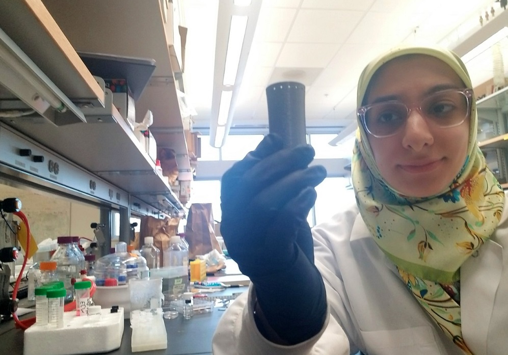
 Together, in conjunction with an interdisciplinary team led by Parinaz Fathi, a fourth-year Bioengineering graduate student in Pan’s laboratories, the investigators were able to develop a solution using a newly popular technique: 3D printing. The use of imaging combined with 3D printing allows for the rapid preparation of completely customized stents.
“The promising part of the study is our ability to image the small intestine using X-ray imaging and get an exact idea of the dimension of the area where to place the stent. This is essential to making the stenting process personalized,” Pan said.
In the future surgeons may be able to place these personalized biodegradable stents at the site of the intestinal perforation in patients with a gastrointestinal tract obstruction or perforation. These stents prevent leakage, provide a surface on which the intestine can heal, and eventually dissolve, preventing further surgeries.
"One of the really exciting things about this work is that it was a perfect opportunity to use engineering to solve a medical problem,” Fathi said. “3D printing is generally very useful, but actually using it to print stents that might one day be clinically used demonstrates the importance of conducting collaborative research with a specific medical problem in mind.”
The team’s study, funded by the Meyer Charitable Trust, progressed rapidly, and researchers identified optimal materials to use for the stent and the best process to produce them. Preliminary studies in pigs have been successful.
Within the next year, the team hopes to conduct more trials in animals and complete full-blown toxicity studies.
“If this works the way we think it will, there is a potential to really improve care and even save lives,” Dr. Rowitz said. “It can change how we manage a very, very difficult problem.”
Categories: Redefining Healthcare
Tags: 3D, Carle, collaborative, engineering, giving, Illinois, of, printing, research, Rowitz, University